Photosynthesis in Higher Plants
Krishna1111
CHAPTER-13 PHOTOSYNTHESIS IN HIGHER PLANTS

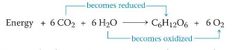
▶️Photosynthesis is an intracellular anabolic process in which green parts of plants synthesise complex organic food (glucose) by using simple inorganic substances such as CO2 (from air) and H2O (from soil) in presence of light and Chl-a with the release of O2 in the atmosphere.
▶️Photosynthesis is a physio-chemical process (in presence of physical energy (light) chemical energy (food, ie., glucose) is formed) or photo-bio-chemical process (rearrangement of molecules by absorption of light energy or conversion of light energy into biochemical energy).
Photosynthesis is redox reaction (Oxidation of H2O & reduction of CO2 takes place).
Importance of Photosynthesis:
1) Photosynthesis is the primary source of food on earth on which all living organisms (heterotrophs) depend.
2) It maintains equilibrium of O2 in the atmosphere.
🔴🔴
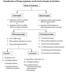
Classification of living organisms on the basis of mode of nutrition:
🔴Mode of Nutrition
▶️Autotrophs
🌲Photoautotrophs
➡️Oxygenic
Ex. 1) Kingdom Plantae
2) Kingdom Monera (BGA)
3) Kingdom Protista (Diatoms & Euglena)
➡️Anoxygenic
Ex. 1) Kingdom Monera
Green sulfure bacteria (Chlorobium) & Purple sulfure bacteria (Chromatium)
🌲Chemoautotrophs
Ex. 1) Nitrifying bacteria (Nitrosomonas)
2) Sulfur bacteria (Thiobacillus)
3) Iron bacteria (Ferrobacillus)
▶️Heterotrophs
First photosynthetic organism on earth was anoxygenic bacteria.
• First oxygenic photosynthetic organisms on earth was cyanobacteria (bluegreen Algae)
• 90% of total photosynthesis is carried out by aquatic plants and 10% is carried out by terrestrial or land plants.
Demonstration
Experiment to show that chlorophyll is essential for photosynthesis.
1) Take a fresh variegated leaf of Croton plant growing in bright sunlight.
2) Place it in boiling water to remove its waxy coating.
3) Then place this leaf in a test tube containing ethanol.
4) This test tube is then put in a beaker filled with hot water till it decolorize.
5) Take out the decolorized leaf & wash it with water to rinse with ethanol.
6) Immerse the leaf in iodine solution. Starch reacts with iodine to give blue colouration.
7) Excess iodine is poured off & the leaf is examined.
8) In a variegated leaf, only green parts (chlorophyll containing) turn blue, while the non green parts remain pale.
9) This shown that chlorophyll is essential for photosynthesis.

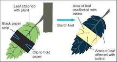
Experiment to show that light is essential for photosynthesis.
1) Take a healthy potted plant and keep it in a dark for 2 to 3 days to destarch it.
2) Select one leaf and partially cover it with black paper.
3) Place this destarched potted plant in light for photosynthesis
4) After 4 to 6 hours, take a leaf that was partially covered with black paper.
5) After carrying out steps of iodine starch test, it was found that entire leaf lamina become blue black except portion of the leaf covered with black paper.
6) Covered leaf area will not show the presence of starch. Thus, the results of this experiment clearly show importance of light in photosynthesis.
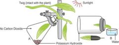
Experiment to show that CO2 is essential for photosynthesis.
[Moll’s half leaf Expt]
1) Take a healthy potted plant and keep it in a dark for about 2 to 3 days. This is done to ensure that the leaves are destarched.
2) Take a test tube or glass bottle containing potassium hydroxide (KOH) solution (or cotton soaked in KOH solution) with a rubber cork. KOH absorbs CO2present in the bottle.
3) The rubber cork is cut into two halves. Select one leaf of destarched potted plant and place it between the two halves of the rubber cork in such a manner that half portion of leaf is in the glass bottle and remaining half leaf is exposed to air.
4) The leaf should remain attached to the potted plant. This set up is then kept in sunlight for 4 to 6 hours.
5) The leaf is then detached from the plant & is removed from bottle.
6) After carrying out steps iodine starch test, it is found that the part of the leaf that was present outside the bottle (exposed to air) shows blue black colour due to formation of starch.
7) The part of leaf enclosed in a glass bottle shows negative test for starch because CO2 was not available to that part as it was absorbed by KOH.
8) This experiment clearly shows importance of CO2 in photosynthesis.
First experiment on photosynthesis
1770:
🔰
Joseph Priestley (1733-1804)
1) Joseph Priestly was the first person who discovered oxygen on August 1, 1744.
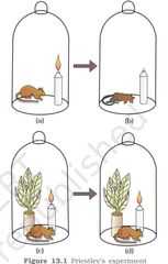
2) He carried out a series of experiments in 1770 using candle, mouse and mint plant that revealed the essential role of air in the growth of plants.
3) He observed that a candle burning in a closed space gets extinguished soon. Similarly, a mouse would soon suffocate in a closed space.
4) He concluded that a burning candle or an animal that breathe the air, but somehow, damage the air.
5) But when he placed a Mint plant in the same bell jar, the mouse stayed alive and the candle continued to burn.
6) Therefore, Priestley hypothesized that plants restore to the air whatever breathing animals and burning candles remove.
7) ▶️He termed the impure air as ‘phlogiston’ and the pure air as ‘dephlogiston’.
1779:
🔰
Jan Ingenhousz (1730-1799):
1) Jan Ingenhousz repeated several times Priestley’s experiment (1779) with an aquatic plant (Hydrilla) in presence and in absence of light.
2) He showed that in the plant that is kept in light, small bubbles were formed around the green parts, while no bubbles appeared in the plant kept in the dark.
3) Later he identified these bubbles to be of oxygen.
4) He concluded that it is only the green part of the plants that can release oxygen (purify air) only in presence of light.
1883:
🔰
T.W.Engelmann (1843-1909):
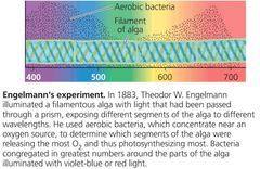
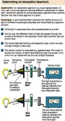
1) T.W. Engelmann in 1883 performed an experiment by using prism, Cladophora and aerobic bacteria.
2) Using a prism, he split light into its spectral components and then he illuminated a green alga, Cladophora placed in a suspension of aerobic bacteria.
3) The bacteria were used to detect the sites of oxygen evolution.
4) He observed that the bacteria accumulated mainly in the region of blue and red light of the split spectrum.
5) A first action spectrum of photosynthesis was thus described.
1854:
🔰
Julius Von Sachs (1832-1897):
1) In 1854 Julius Von Sachs showed that glucose is produced when plants grow.
2) He also showed that glucose is stored as starch.
3) His later studies showed that green substance in plants (chlorophyll as we know it now) is located in special bodies (later called chloroplast) within the plant cells.
By the middle of the nineteenth century
By the middle of the nineteenth century the key features of plant photosynthesis were know, namely, that plants could use light energy to make carbohydrates from CO2 and H2O.
The empirical equation representing the total process of photosynthesis for oxygen evolving organisms was the understood as :
CO2 + H2O →Light → [CH2O] + O2
Where [CH2O] represented a carbohydrate (glucose)
1930:
🔰
Cornelius Van Viel (1897-1985:
1) A milestone contribution to the understanding of photosynthesis was that made by microbiologists C.V. Niel (1930) on the basis of his study on purple & green sulfur bacteria.
2) He demonstrated that photosynthesis is essentially a light dependent reaction in which hydrogen from a suitable oxidisable compound (Hydrogen donor) reduces carbon dioxide to carbohydrates.
2H2 A + CO2 →Light → 2 A + CH2O + H2O
3) In green plants H2O is the hydrogen donor and is oxidised to O2.
4) Some organisms such as purple and green sulfur bacteria do not release O2during photosynthesis.
5) Purple and green sulfur bacteria use H2S as the hydrogen donor. The ‘oxidation’ product is sulfur or sulfate depending on the organism and not O2.
6) Hence, he inferred that the O2 evolved by the green plant comes from H2O, not from carbon dioxide.7) This was later proved by using radioisotopes of oxygen (H2O18) (Ruben & Kamen).
1937:
Robert Hill / (Robin Hill) (1899-1991) :
1) Robert Hill (1937) performed an experiment in which he suspended isolated chloroplasts from spinach leaves in water without CO2.2) He then added ferric salt or (Dichloro-phenol Indophenol, a blue dye) as hydrogen acceptor (electron acceptor) and the suspension was exposed to sunlight.
3) He observed that ferric salt is reduced to ferrous (DCPIP turn into colourless) and O2 bubbles evolve
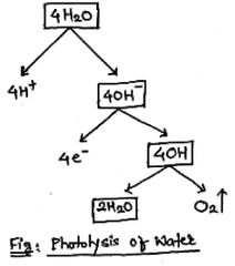
4) Thus, he clearly proved that H2O splits. This is called photolysis of water or Hill reaction and is represented as follows
2H2O + 2A→Sunlight, Chlorophyll→ 2 AH2 + O2 H2O
Here ‘A’ is unknown hydrogen acceptor present in chloroplast.
Hill reaction: Isolated chloroplasts can bring about dissociation of water in presence of light and suitable ‘H’ acceptor (Robert Reagent).�
1) Ruben and kamen in 1941 used heavy, isotope of oxygen, i.e., O18 and proved that the source of O2 evolved during photosynthesis is water.
1) Dr. Arnon in 1954 discovered that the unknown hydrogen acceptor (A-in Hill reaction) is a Co-enzyme, Nicotinamide Adenine Dinucleotide Phosphate (NADP).
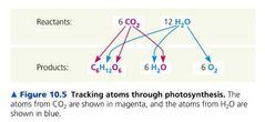
Tracking atoms through photosynthesis
WHERE DOES PHOTOSYNTHESIS TAKE PLACE?
‘the green leaf’ or ‘in the chloroplasts’,
Q) Can you name some other parts where you think photosynthesis may occur?
Answer:
Stem:- Opuntia, Euphorbia & Cactus (Phylloclade);
Asparagus(Cladode)
Root:- Epiphytic roots of Epiphytes (Orchids); Adventitious roots of Tinospora & Trapa.
Phyllode :- Australian Acacia.
Calyx, Tender fruits & Tendril are also photosynthetic
Chloroplast
Shape
Size
Ultrastructure
Chloroplast is a type of plastid.
Shape:- Variable ; star-shaped or stellate (Zygnema), cup-shaped (Chlamydomonas), spiral (Spirogyra); in higher plants it is lens-shaped or biconvex.
Size:- In higher plants - width 1 to 2 ìm & length 4 to 6 ìm.
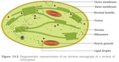
Ultrastructure:-
1) Chloroplast consists of two membranes, i.e., outer membrane & inner membrane together called as 💡peristromium or envelope.
2) In between outer membrane & inner membrane there is space called as 💡Periplastidial space.
3) Inside peristromium a space is present which is filled with proteinous matrix called as 💡stroma.
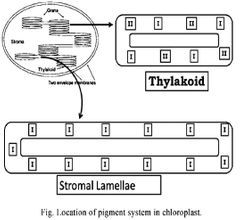
4) Inside stroma a complex network of membrane is present. The unit of internal complex network of membrane is 💡thylakoid.
5) Thylakoids are placed one above one like stacks of coin & form a structure called 💡grana.
6) Two grana are connected with each other by a lamellae called as 💡intergranal lamellae or stromal lamellae or FRET membrane.
7) Stroma consist of single, double stranded, circular, closed & naked DNA as a genetic material called as 💡plastidome.
8) 70S ribosomes are also present.
9) Lipid droplets (Plastoglobules) & starch grains are also present.
10) All enzymes necessary for CO2 fixation is also present in stroma.
Alignment of chloroplasts
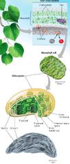
The mesophyll cells in the leaves, have a large number of chloroplasts.
Q) Usually the chloroplasts align themselves along the walls of the mesophyll cells, why?
Q) When do you think the chloroplasts will be aligned with their flat surfaces parallel to the walls?
Q) When would they be perpendicular to the incident light?
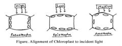
Usually the chloroplasts alignthemselves along the walls of the mesophyll cells, such that they get the optimum quantity of the incident light. And for easy diffusion of gases.
Answer:
Usually chloroplasts align themselves along the walls of mesophyll cells.
When light intensity is high, chloroplasts align themselves along the walls that are parallel to the incident light (Parastrophe).
When light intensity is low, chloroplasts tend to align along the cell walls that are perpendicular to the incident light in order to obtain maximum light (Epistrophe).
When light intensity is optimum (medium) they align randomly (Apostrophe)
How many types of pigments are involved in photosynthesis?
🔰A chromatographic separation of the leaf pigments shows that the colour that we see in leaves is not due to a single pigment but due to four pigments :
🔰Chlorophyll - a (bright or blue green in the chromatogram),
Chlorophyll -b (yellow green),
Xanthophylls (yellow) and
Carotenes (orange) [carotenoids (yellow - orange)]
🔰) Pigments are substances that have an ability to absorb light, at specific wavelengths.
▶️) All those pigments involved in photosynthesis are called as photosynthetic pigments.
1⃣Chlorophyll (Green)
Chl-a ,
, Chl-b, Chl-c, Chl-d, Chl-e, Bacteriochlorophyll, Bacterioviridin
2⃣Carotenoids
Carotenes (Orange)
Xanthophylls (Yellow)
3⃣Phycobillins
Phycocyanin (Blue)
Phycoerytrin (Red)
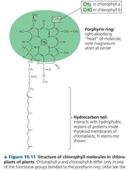
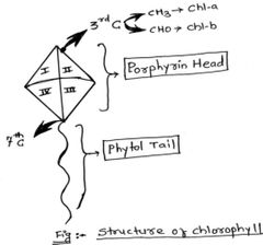
Chlorophyll:
Chlorophyll pigments are insoluble in water
There are major seven types of chlorophyll pigments.
Chlorophyll has kite-like or tad-pole like structure.
It has two parts, i.e., Head and Tail.
▶️Chemically head region is porphyrin ring (cyclic tetrapyrrole ring joined together by –CH= bridge) with ‘Mg’ atom at its center.
▶️ Tail is simple hydrocarbon chain called as phytol chain. ( chemically acyclic diterpene alcohol).
Tail is joined with head region at 7th carbon of 4th pyrrole ring.
In Chl-a there is CH3 and in Chl-b there is CHO group at 3rd carbon atom of 2nd pyrrole ring.
Function: Chlorophyll acts as a reaction center. It is pigment involved in photosynthesis.
⚠️Chlorophyll a
C55 H72 O5 N4 Mg
There are major seven types of chlorophyll pigments.
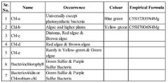
Chl-a
Universally except photosynthetic bacteria
Blue green
C55H72O5N4Mg
Chl-b
Algae and higher plants
Yellow green
C55H70O6N4Mg
Chl-c
Diatoms, Red algae & Brown algae
Chl-d
Red algae & Brown algae
Chl-e
Rarely in Yellow-green & Green algae
Bacteriochlorophyll
Green Sulfur & Purple Sulfur Bacteria
Bacterioviridin or Chlorobium chl.
Green Sulfur & Purple Sulfur Bacteria
Biosynthesis of chlorophyll
a) In plants Glycine and succinyl Co-A from Krebs cycle are first precursor for chlorophyll synthesis.
b) After a series of reaction a compound protochlorophyllide is formed.
c) It is pale yellow in colour. Light acts as a catalyst for the synthesis of mature chlorophyll.
d) Light induced reduction of protochlorophyllide gives chlorophyllide.
e) To chlorophyllide, pytol gets attached and Chlorophyll is formed (by esterification)
f) Chlorophyll synthesis is plants require light, N, Mg, S, Fe, K, Ca.g) Light is essential for to synthesis of chlorophyll in Angiosperms only. Exceptions are Citrus of Nelumbo. Algae, Bryophytes, Pteridophytes and Gymnosperms can synthesise chlorophyll even in absence of light.
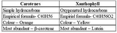
1) Carotenoid pigments are insoluble in water. They are soluble in organic solvents.
2) They are derived lipid compounds.
3) They are derivatives of tetraterpenes, i.e., they are produced from 8 isoprene units [CH2=C(CH3)-CH=CH2].
4) There are two types of carotenoids, i.e., carotenes and xanthophylls
Function/ Importance:
a) They act as accessory or antenna pigments.
b) They prevent solarization and photo-oxidation of chlorophyll hence called as shield pigment.
1) Phycobillins are soluble in water.
2) They have open tetrayrrole structure lacking ‘Mg’ atom.
3) There are three types of phycobillins, i.e.,
Allophycocyanin,
phycocyanin and
phycoerythrin.
4) Allophycocyanin & Phycocyanin is blue coloured pigment found only in BGA while phycoerythrin is red couloured pigment present only in Red algae.
Nature of Light
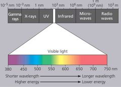
) Light is a visible part of electromagnetic spectrum.
⚠️) Only visible light (400nm - 700 nm) is used in photosynthesis. therefore, it is referred as photosynthetically Active Radiations (PAR)
) Light energy shows a dual nature. Light propagates through space by waves. Light emitted from a source behave as it is made up of discrete unit or particles called photons.
) The energy carried by a photon is called quantum (pl.quanta)
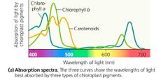
Absorption spectrum is a graph showing amount of absorption of light energy of different wavelength by photosynthetic pigments.
Q. Which is the most abundant plant pigment in the world?
Q. From Fig. (Graph showing absorption spectrum of Chl-a, Chl-b and Carotenoids) can you determine the wavelength (colour of light) at which chl-a shows the maximum absorption?
Q. Does it show another absorption peak at any other wavelengths too?
Q. If yes, which one?
Ans. Chlorophyll-a is the most abundant plant pigment in the world.
Ans. Chlorophyll-a shows maximum absorption in 430 nm (Blue colour of light).
Ans. Yes.
Ans. 662 nm (Red colour of light).
Absorption spectrum of photosynthetic pigment
) These graphs (absorption spectrum of photosynthetic pigment and action spectrum of photosynthesis), show that most of the photosynthesis takes place in the blue and red regions of the spectrum; some photosynthesis does takes place at the other wavelengths of the visible spectrum.
) Let us see how this happens. Though chlorophyll is the major pigment responsible for trapping of light, other thylakoid pigments like chlorophyll-b, xanthophylls and carotenes, which are called accessory pigments, also absorb light and transfer the energy to chlorophyll-a.
) Indeed, they not only enable a wider range of wavelength of incoming light to be utilized for photosynthesis but also protect chlorophyll-a from photo-oxidation.
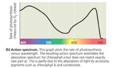
An action spectrum is the graph showing relative rates of photosynthesis at different wavelength of light.
It shows which wavelength of light is most effectively used in a specific chemical reaction.
How, we can conclude that chlorophyll-a is the chief pigment associated with photosynthesis.
By looking at absorption spectrum of chlorophyll-a and action spectrum of photosynthesis, one can clearly see that the wavelength at which there is maximum absorption of chlorophyll-a, i.e., in the blue and red regions, also shows higher rate of photosynthesis.
) Hence, we can conclude that chlorophyll-a is the chief pigment associated with photosynthesis.
Q. By looking at figure (Graph showing action spectrum of photosynthesis superimposed on absorption spectrum of chlorophyll-a) can you say that there is a complete one-to-one overlap between the absorption spectrum of chl-a and the action spectrum of photosynthesis?
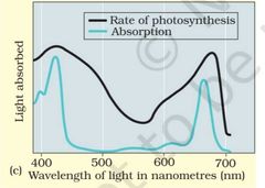
Ans. No, there is no complete one to one overlap between the absorption spectrum of chlorophyll-a and the action spectrum of photosynthesis because at certain points action is more than absorption because wavelengths not absorbed by chlorophyll-a are absorbed by accessory pigments and transferred to reaction center (chlorophyll-a).
Emerson’s Experiments
Evidence for two pigment systems
Emerson’s first experiment
(Red drop effect):-
Emerson’s second experiment (Enhancement effect) :-
Emerson’s first experiment
(Red drop effect):-
Emerson’s second experiment (Enhancement effect) :-
Two pigment system
The discovery of Emerson’s effect clearly indicates that two separate group of pigments are involved in photosynthesis.
One pigment complex is driven by higher wavelength while other is driven by lower wavelength
) They gave the concept of two photo system or two pigment system.
) These photosystems are present (membrane of thylakoid, and stromal lamellae) in internal complex network of membrane and from morphologically distinct physical entities called quantasome.
) These were discovered by Park and Biggins the first time in 1964.
Quantasome is a smallest group of collaborating pigment molecules necssary to affect a photochemical act, i.e., absorption of light energy and transfer of absorbed energy to respective reaction center where it can promote release of an electron.
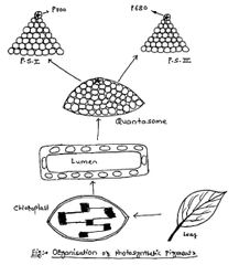
Quantasome = Photo System = Pigment System
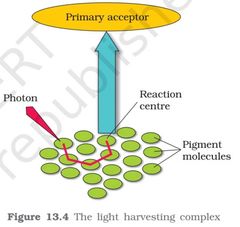
A Photosystem: A Reaction-Center Complex Associated with Light-Harvesting Complexes
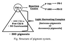
In environment of the thylakoid membrane, chlorophyll molecules are organized along with other small organic molecules and proteins into complexes called photosystems.
Each photosystem consists of Light Harvesting Complex (LHC) and a Reaction center
The LHC are made up of hundreds of pigment molecules bound to proteins.
Each photosystem has all the pigments(except one molecule of chlorophyll a) forming a light harvesting system also called antennae.
These pigments help to make photosynthesis more efficient by absorbing different wavelengths of light.
) LHC is composed of about 200 Chl-a, few Chl-b and 50 carotenoids.
) Reaction center is single specific chlorophyll molecule.
) PS-I has P-700 and PS-II has P-680 as a reaction center.
The thylakoid membrane is populated by two types of photosystems that cooperate in the light reactions of photosynthesis. They are
They are called photosystem II (PS II) and photosystem I (PS I).
(They were named in order of their discovery, but photosystem II functions first in the light reactions.)
Each has a characteristic reaction-center complex—a particular kind of primary electron acceptor next to a special pair of chlorophyll a molecules associated with specific proteins.
These two pigments, P680 and P700, are nearly identical chlorophyll a molecules. However, their association with different proteins in the thylakoid membrane affects the electron distribution in the two pigments and accounts for the slight differences in their light-absorbing properties.
The reaction-center chlorophyll a of photosystem II is known as P680 because this pigment is best at absorbing light having a wavelength of 680 nm (in the red part of the spectrum).
The chlorophyll a at the reaction-center complex of photosystem I is called P700 because it most effectively absorbs light of wavelength 700 nm (in the far-red part of the spectrum).
Mechanism of Photosynthesis:
) The entire mechanism of photosynthesis takes place in chloroplast.
) It is divided into two phases, i.e., light reaction and dark reaction.
1) It is light dependent phase of photosynthesis.
2) Light reaction takes place in grana portion of the chloroplast.
3) It is photo-biochemical process and responsible for the formation of assimilatory power, i.e., NADPH2 (reducing power) and ATP (energy donor).
4) ATP and NADPH2 are high-energy chemical intermediates.
5) Two important events, i.e., photo-phosphorylation and photolysis of water take place during light reaction. It is also known as Hill reaction.
Light reaction consists of five steps:
[1] Absorption of light energy by photosyntheticpigments.
[2] Transfer of light energy by photosynthetic pigments to respective reaction center.
[3] Excitation of Chl–a (Reaction center) & ejection of electron.
[4] Photophosphorylation and
[5] Photolysis of water.
▶️Synthesis of ATP from ADP & iP is called as phosphorylation. It is endothermic reaction and requires energy. Since this energy comes from light it is called as photophosphorylation.
) Dr. Arnon first suggested that there are two types of photophosphorylation, i.e., Cyclic and Non-cyclic photophosphorylation.
Old Concept
Kinetic energy of electron is responsible for phosphorylation
mistake no enzyme
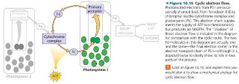
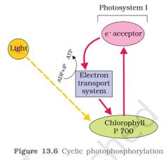
(A) Cyclic photophosphorylation
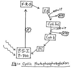
Cyclic photophosphorylation takes place in stromal lamellae.
2) Only one pigment system (P.S. 1) is involved in cyclic photophosphorylation.
3) All pigments in P.S.I absorb light energy and transfer absorbed light energy to reaction center, i.e., P-700 by resonance.
4) Reaction center undergo excitation and ejects its electron with extra amount of energy.
5) Ejected electron is accepted by Primary electron acceptor of P.S.I, i.e., FRS (Ferredoxin Reducing Substance).
6) The movement of electron from P-700 to primary electron acceptor (FRS) is called as uphill movement.
7) From FRS electron is transferred through electron transport system containing various intermediate electron carriers.
8) They are Ferredoxin (Fd), Cytochrome complex (Cyt.b6 & Cyt-f) & Plastocyanin (PC).
9) Finally, deenergized electron comes back to its original position, i.e., Chl-a (P-700).
10) This movement of electron from FRS to P-700 is downhill (electron loses its energy from each transfer).
11) Various electron carriers are arranged according to their redox potential scale (increasing affinity towards electron).
12)Redox Potential is the measure of the tendency of a chemical molecule to acquire electrons and thereby be reduced. It is also called as oxidation-reduction potential.
13) The electron is not used up as it passes through the various electron carriers & finally come back to its original position, i.e., P-700 in a cyclic manner.
14) At two places during the transfer of electrons, i.e., from Ferredoxin (Fd) to Cyt.b6 & from Cyt.b6 to Cyt.-f enough energy is released which is used for the synthesis of ATP.
15) Thus, cyclic movement of electron is coupled with the synthesis of ATP, it is called as cyclic photophosphorylation.
16) The cyclic flow of electron hence, results only in the synthesis of ATP but not NADPH2.
Cyclic photophosphorylation also occurs when only light of wavelengths beyond 680 nm are available for excitation.
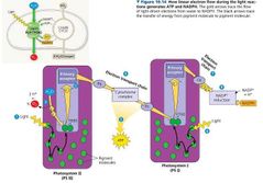
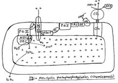
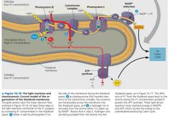
(B) Non-Cyclic photophosphorylation:
Non-cyclic photophosphorylation takes place in granal thylakoid.
2) Both pigment systems, i.e., PS-I & PS-II are involved in non-cyclic photophosphorylation.
3) All pigments in P.S.-1 absorb light energy and transfer absorbed light energy to reaction center, i.e., P-700 by resonance.
4) Reaction center undergo excitation and ejects its electron with extra amount of energy. A positive charge is developed on P-700 (electron deficiency is created).
5) Ejected electron is accepted by primary electron acceptor of PS-I, i.e., FRS (Ferredoxin Reducing Substance).
6) The movement of electron from P-700 to primary electron acceptor is called as uphillmovement.
7) From FRS electron is transferred to Ferredoxin (Fd).
8) From Ferredoxin this electron is transferred to NADP via NADP-Reductase enzyme which is located towards stroma side of the thylakoid.
9) Movement of electron from FRS to NADP is downhill.
10) Electron deficiency created in PS-I which is fulfilled by electron coming from PS-II.
11) All pigments of PS-2 absorb light energy and transfer its energy to reaction center, i.e., P-680 by resonance.
12) P-680 absorb 680 nm wavelength of red-light causing electrons to become excited and jump into an orbit farther from the atomic nucleus.
13) Ejected electron is first accepted by primary electron acceptor, i.e., Coenzyme-Q (Co-Q).
14) This movement of electron from P-680 to Co-Q is uphill movement. 15) From Co-Q electron is transferred through various electron carriers including Plastoquinone (PQ), Cytochorome complex (Cyt.b6 & Cyt-f) & Plastocyanin (PC).
16) From Plastocyanin de-energized electron is transferred to PS-I. This movement of electron is downhill.
17) At one place during the transfer of electrons, i.e., in between Cyt.b6 & Cyt.-f enough energy is released which is used for the synthesis of ATP.
18) Electron deficiency of PS-I is fulfilled by electron of PS-II but electron deficiency is now created in PS-II.
19) We need to emphasize here that the water splitting complex is associated with the PSII, which itself is physically located on the inner side or the membrane of the thylakoid.
20) This complex is responsible for photolysis of water
21) Ionized PS-II acts as a strong oxidant and brings about photolysis of water.
22) Water molecules split into electron, proton and oxygen.
23) Electrons are accepted by PS-II. Thus, water acts as an ultimate source of electron in photosynthesis. 24) Protons are accepted by NADP-- and get converted into NADPH2.
25) Oxygen gets diffused out from thylakoid.
Electron ejected from PS-II & PS-I never come back to its original position as it is accepted by NADP. Therefore, this movement of electron is called as non-cyclic movement and resulting synthesis of ATP is called as non-cyclic photophosphorylation.
Non-cyclic photophosphorylation is very significant process because ATP and NADPH2 are formed during this process.
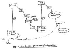
Z-scheme
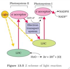
Non-cyclic photophosphorylation.
This whole scheme of transfer of electrons starting from the PS II uphill to the acceptor down the electron transport chain to P. S. I, excitation of electrons, transfer to another acceptor and finally downhill to NADP reducing it into NADPH2 is called Z-scheme due to characteristic shape.
This shape is formed when all the electron carriers are placed in a sequence on a redox potential scale. 29) Non-cyclic photophosphorylation is very significant process because ATP and NADPH2 are formed during this process�
Difference between Cyclic and Non-cyclic photophosphorylation.
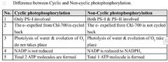
Cyclic photophosphorylation
1. Only PS–I involved
2. The e- expelled from Chl-700 is cycled back
3. Photolysis of water & evolution of O2 do not takes place
4. NADP is not reduced
5. Total 2 ATP molecules are formed
Non-Cyclic photophosphorylation
1. Both PS–I & PS–II involved
2. The e- expelled from Chl-700 is not cycled back
3. Photolysis of water & evolution of O2 takes place
4. NADP is reduced to NADPH2
5. Total 1 ATP molecule is formed
Prof. Peter Mitchell proposed Chemiosmotic theory in 1961 to explain mechanism of ATP synthesis in mitochondria and chloroplast.
2) According to him ATP synthesis, i.e., phosphorylation is purely an enzymatic process.
3) ATP synthase enzyme is responsible for the ATP synthesis.
4) According to chemiosmotic theory, chemiosmosis, i.e., movement of hydrogen ions from the region of higher concentration to lower concentration release energy which drives the process of phosphorylation.
5) Like in respiration, in photosynthesis too, ATP synthesis is linked to development of a proton gradient across a membrane.
6) Chemiosmosis is coupled with phosphorylation. Therefore, chemiosmotic theory is also known as coupling theory.
Chemiosmosis requires
semipermeable membrane,
ATP synthase enzyme and
H+ ion concentration gradient.
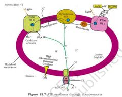
Prof. Peter Mitchell proposed three causes for the development of proton gradient inside thylakoid lumen.
(a) Since splitting of the water molecules take place on the inner side of the membrane, the protons or hydrogen ions that are produced by the splitting of water accumulate within the lumen of thylakoid.
(b) As electrons move through the photosystems (PS-II to PS-I), protons are transported across the membrane (from stroma to lumen). This happens because the primary acceptor of electron (CO-Q) which is located towards the outer side of the membrane transfers its electron not to an electron carrier but to an H carrier (PQ). Hence, thismolecule removes a proton from the stroma while transporting an electron. When this molecule passes on its electron to the electron carrier (Cyt. b6-Cyt. f complex) on the inner side of the membrane, the proton is released in to the inner side or the lumen side of the membrane.
(c) The NADP reductase enzyme is located on the stoma side of the membrane. Along with electrons that come from the acceptor o electrons of PS I, protons are necessary for the reduction of NADP to NADPH2. These protons are also removed from the stroma.
Hence, within the chloroplast, protons in the stroma decrease in number, while in the lumen there is accumulation of protons. This creates a proton gradient across the thylakoid membrane as well as a measurable decrease in pH in the lumen.
H+ Concentration gradient is important because
This gradient is important because it is the breakdown of this gradient that leads to the synthesis of ATP.
) The gradient is broken down due to the movement of protons (chemiosmosis) across the membrane (from lumen) to the stroma through the transmembrane channel of the CF0 of the ATP synthase and release enough energy.
) This kinetic energy of protons is utilized by ATP synthase enzyme for phosphorylation
ATP synthase (CF0-CF1) enzyme has two parts:
one called as CF0 is embedded in the membranes (an integral membrane protein complex) and forms a transmembrane channel that carries out facilitated diffusion (protons channel) of protons across the membrane.
) The other portion is called as CF1 and protrudes on the outer surface of the thylakoid membrane (peripheral membrane protein complex) on the side that faces the stroma. CF1 act as actual catalytic site for ATP synthesis.
) The breakdown of the gradient provides enough energy to cause a conformational change in the CF1 particle of the ATP synthase, which makes the enzyme synthesize several molecules of energy-packed ATP.
) Thus, during chemiosmosis, concentration gradient broken-down and kinetic energy of protons is released. ATP synthase enzyme channelize movement of protons and use released energy effectively for the synthesis of ATP.
) Hence ATP synthase (FO-F1) is called as Coupling Factor (CFO-CF1).
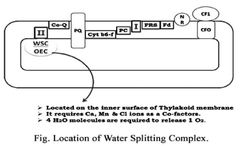
Photolysis of Water:
1) Photolysis of water takes place on the inner surface of thylakoid membrane.
2) Unknown protein complex called as water splitting complex or oxygen evolving complex is associated with PS II.
3) It requires Ca, Mn & Cl ions as a Co-factors.
4) It uses light energy to split water.
5) PS II acts as a strong oxidant in photolysis of water.
6) Four water molecules are required for Water Splitting Complex or Oxygen Evolving Complex to release one O2
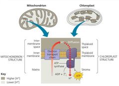
A Comparison of Chemiosmosis in Chloroplasts and Mitochondria
1) The products of light reaction are ATP, NADPH2 and O2 . Of these O2 diffuses out of the chloroplast while ATP and NADPH2 are used to drive the processes leading to the synthesis of food, more accurately, sugars.
2) This is biosynthetic phase of photosynthesis.
3) This process does not directly depend on the presence of light but is dependent on the products of the light reaction, i.e., ATP and NADPH2, besides CO2 and H2 O.
4) You may wonder how this could be verified; it is simple immediately after light becomes unavailable, the biosynthetic process continues for some time and then stops. If then, light is made available, the synthesis starts again.
5) Dark reaction takes place in stroma portion of the chloroplast.
6) It is also called as Blackmans reaction.
Q. Can we, hence, say that calling the biosynthetic phase as the dark reaction is a misnomer. Discuss this amongst yourselves.
Ans. Dark reaction doesn’t depend on light but it depends on the products of light reaction like ATP and NADPH2. In the absence of light, the light reaction can not take place. However, it is observed that after sometime biosynthetic phase continues for a while. This shows that dark reaction is indirectly dependent on light and requires the products of light reaction to proceed. Hence, using the term dark reaction to describe the biosynthetic phase is indeed, misleading.
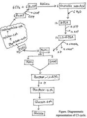
Melvin Calvin and his co-workers (Benson and Basham) discovered the pathway of glucose synthesis in 1954 (Nobel Prize 1961).
) Calvin and his co-workers showed that biochemical reactions take place in a cyclic manner hence this biosynthetic pathway is called as Calvin cycle or Calvin-Benson cycle.
) First stable product formed after CO2 fixation is 3 Carbon containing compound, i.e., PGA (phosphogylceric acid). Therefore, it is also called as C-3 cycle.
) It is also called as Photosynthetic Carbon Reduction cycle (PCR) or Carbon Assimilation Cycle (CAC).
🔰We saw earlier that CO2 is combined with H2O to produce (CH2O)n or sugars. It was of interest to scientists to find out how this reaction proceeded, or rather what was the first product formed when CO2 is taken into a reaction or fixed. Just after world war II, among the several efforts to put radioisotopes to beneficial use, the work of Melvin Calvin is exemplary. The use of radioactive 14C by him in algal photosynthesis studies led to the discovery that the first CO2 fixation product was a 3-carbon organic acid. He also contributed to working out the complete biosynthetic pathway; hence it was called Calvin cycle after him.
Melvin Calvin and his co-workers (Benson and Basham) used following test material and techniques to discover glucose synthetic pathway.Test Material:
(1) Chlorella & Scenedesmus (Unicellular green algae)
(2) Radioactive isotope of Carbon 14CO2
Techniques:
(1) Paper chromatography (To separate the intermediates)
(2) Autoradiography (To find out compounds with 14CO2 )
The Primary Acceptor of CO2
How many carbon atoms would a molecule have which after accepting (fixing) CO2, would have 3 carbons (of PGA)?
The studies very unexpectedly showed that the acceptor molecule was a 5-carbon ketose sugar – ribulose bisphosphate (RuBP).
Scientists also believed that since the first product was a C3 acid, the primary acceptor would be a 2-carbon compound; they spent many years trying to identify a 2-carbon compound before they discovered the 5-carbon RuBP.
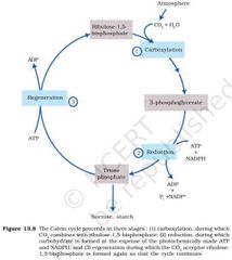
Calvin Cycle
Three major steps
Calvin described glucose synthetic pathway in three major steps, i.e., Carboxylation, Reduction and
Regeneration.
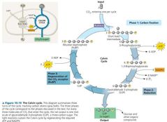
a) Carboxylation:-
1) Carboxylation is the fixation of CO2 into a stable organic intermediate.
2) Carboxylation is the most crucial step of the Calvin cycle where CO2 is utilized for thecarboxylation of RUBP.
3) Atmospheric CO2 is first accepted by a keto-pentose sugar, i.e., Ribulose,l-5,bisphosphate (RUBP) in presence of enzyme RUBP Carboxylase.
4) After carboxylation of RUBP it gets converted into two molecules of 3-carbon compound called 3-phosphoglyeric acid (PGA).
5) Since the enzyme also has an oxygenation activity it would be more correct to call itRUBP carboxylase-oxygenase or RuBisCO.

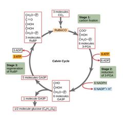
b) Reduction:-
1) These are a series of reactions that lead to the formation of glucose. The stpes involve utilisation of 2 molecules of ATP for phosphorylation and two of NADPH2 for reduction per CO2 molecule fixed.2) Thus, two phosphoglyceric acid (3-PGA) molecules are phosphorylated by using two ATP to produce two molecules of 1, 3 di-phosphoglyceric acid (1,3-diPGA). 3) These two 1,3-diPGA molecules then reduced by using two molecules of NADPH2 to produce two phospho-glyceraldehyde (PGAL). 4) The fixation of six molecules of CO2 , six turns of cycle are required for the formation of one molecule glucose from the pathway. 5) Thus, total 12 molecules of PGAL are formed after fixation of six CO2 in six turns of C-3 cycle. 6) 1/6 part of PGAL i.e. out of 12 molecules, 2 molecules of PGAL are used for synthesis of glucose and remaining (5/6 part) PGAL molecules are diverted for regeneration of RUDP. 7) Out of these two molecules, one PGAL is isomerized into dihydroxyacetone phosphate(DHAP). 8) PGAL and DHAP combine together to form one molecule of fructose-1, 6-diphosphate. 9) Fructose-1,6-diphosphate is converted into glucose-6-phosphate after dephosphorylation and isomerization. 10) Finally, Glucose-6-phosphate is converted into Glucose by dephosphorylation�

c) Regeneration:-
1) Regeneration of the CO2 acceptor molecule RuBP is crucial if the cycle is to continue uninterrupted. The regeneration steps require one ATP for phosphorylation to form RuBP.
2) In six turns of this cycle total six ATP molecules are required in regeneration step.
3) Hence, for every CO2 molecule entering the Calvin cycle, 3 molecules of ATP and 2 NADPH2 are required.
🔴It is probably to meet this difference in number of ATP andNADPH2 used in the dark reaction that the cyclic phosphorylation takes place.�
Hence, for every CO2 molecule entering the Calvin cycle, 3 molecules of ATP and 2 NADPH2 are required. It is probably to meet this difference in number of ATP and NADPH2 used in the dark reaction that the cyclic phosphorylation takes place.
Calvin cycle occurs universally in all photosynthetic autotrophs. To make one molecule of glucose 6 turns of the cycle are required.
5) Total 18 ATPs and 12 NADPH2 molecules are required to synthesize one glucose molecule.
Work out how ATP and NADPH2 molecules will be required to make one molecule of glucose through the Calvin cycle.
CO2. ATP. NADPH2
01. 03. 02
03. 09. 06
06. 18. 12
what goes in and what comes out of the Calvin cycle
In. Out
Six CO2. One glucose
18 ATP. 18 ADP
12 NADPH. 12 NADP
Photorespiration:
[C-2 cycle/ Glycolate Metabolism/ Photosynthetic Carbon Oxidation cycle (PCO)]
Light dependent O2 uptake and release of CO2 is called as photorespiration.
first step pf the Calvin pathway, i.e., CO2 fixation step. This is the reaction where RuBP combines with CO2 to form 2 molecules of 3PGA, that is catalysed by RuBisCO.
RuBisCO that is the most abundant enzyme in the world is characterised by the fact its active site can bind to both CO2 and O2 – hence the name (RibuloseBisphosphate Carboxylase Oxygenase) . 5) RuBisCO has much greater affinity for CO2 when the CO2 :O2 is nearly equal than for O2. This binding is competitive. It is the relative concentration of O2 and CO2 that determine which of the two will bind to the enzyme. 6) Thus, RuBisCO has dual nature of its catalytic activity (it can bind with both CO2 and O2). It is thermolabile enzyme (it changes its activity according to temperature).
In C3 plants some O2 does bind to RuBisCO, and hence CO2 fixation is decreased. Here the RuBP instead of being converted to two molecules of PGA binds with O2 to form one molecule of phosphoglycerate (3 carbon) and phosphoglycolate (2 carbon) in a pathway called photorespiration.
) In the photorespiratory pathway, there is ⚠️neither synthesis of sugars, nor of ATP. Rather it results in the release of CO2 with the utilisation of ATP. In the photorespiratory pathway there is no synthesis of ATP or NADPH. The biological function of photorespiration is not known yet.
) Photorespiration was first observed by Decker & Tio (1959) while studying the mechanism of photosynthesis in Tobacco leaves. Detail pathway was described by Krotkov et al., in 1963.
Photorespiration pathway involves three cell organelles, i.e.,
Photorespiration pathway involves three cell organelles, i.e.,
cloroplast,
perexisome and
mitochondria
Mechanism of photorespiratory carbon oxidation cycle
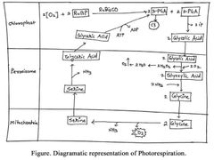
▶️At high temperature, high light intensity and low CO2 concentration RuBisCO acts as oxygenase. Thus, oxidation of RUBP by O2 takes place.
) It results in the formation of one molecule of 2-C compound, phosphoglycolate & one molecule of 3PGA.
) 3-PGA enters Calvin cycle while the 2-phosphoglycolate gets dephosphorylated to form glycolate within the chloroplast.
) The Glycolate then gets enters into peroxisomes. Here it is oxidised again to glyoxylate & then gets converted into a glycine after amination.
) Glycine enters in mitochondria and two molecules of glycine combine to form serine (3C) after decarboxylation and deamination.
) The serine is taken up by peroxisome and gets converted into glycerate after deamination.
) The glycerate enters the chloroplast, gets phosphorylated to form PGA and enters the Calvin cycle.
) Thus, 75% carbon is successfully recovered but 25 % carbon is lost from already fixed carbon.
▶️No. of cell organelles involved.
03
▶️Total No. of O2 consumed.
04
▶️Net O2 consumed.
03
▶️Net ATP consumed.
01
▶️No. of amino acids formed.
Glycine and Serine
▶️Site of oxygenation
Chloroplast & Peroxisome
▶️Site of amination
Peroxisome
▶️Site of deamination
Mitochondria & Peroxisome
▶️Site of decarboxylation
Mitochondria
The Warburg effect is the decrease in the rate of photosynthesis by high oxygen concentrations. Oxygen is a competitive inhibitor of the carbondioxide fixation by RuBisCO. Furthermore, oxygen promotes photorespiration which reduces photosynthetic yield.
The C-4 Pathway:
CO2 Concentrating Mechanism or
Co-operative Photosynthesis or
HSK Pathway or
Hatch and Slack Pathway]
Kortshak, Harrt & Burr in 1965 first time reported alternative method of CO2 fixation while studying the mechanism of photosynthesis in Sugarcane leaves.
) Later M. D. Hatch & C. R. Slack (1970) described entire pathway in a cyclic manner called as Hatch & Slack cycle or HSK cycle.
) First Stable product formed after CO2 fixation is 4 carbon containing Oxalo Acetic Acid. Therefore, it is also called as C-4 Cycle.
) This type of adaptive pathway found in both dicot and monocot plants having tropical origin. They are called as C-4 plants.
Plants that are adapted to dry tropical regions
Plants that are adapted to dry tropical regions have the C4 pathway. Though these plants have the C4 oxaloacetic acid as the first CO2 fixation product they use the C3 pathway or the Calvin cycle as the main biosynthetic pathway.
C4 plants are special: They have a special type of leaf anatomy, they tolerate higher temperatures, they show a response to high light intensities, they lack a process called photorespiration and have greater productivity of biomass.
Adaptations
Which plants?
Why?
▶️Which plants⚠️
Plants that are adapted to dry tropical regions
Dicot plants: Amaranthus, Atriplex Monocot plants: Maize, Sugarcane, Jowar ( Poace) sorghum
▶️Why
To Avoid photorespiration .
To increase productivity.
Study vertical sections of leaves, one of a C3 plant and the other of a C4 plant.
Do you notice the differences?
Do both have the same types of mesophylls?
Do they have similar cells around the vascular bundle sheath?
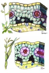
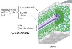
‘Kranz’ anatomy
The particularly large cells around the vascular bundles of the C4 pathway plants are called bundle sheath cells, and the leaves which have such anatomy are said to have ‘Kranz’ anatomy. ‘Kranz’ means ‘wreath’ and is a reflection of the arrangement of cells.
▶️ Homogeneous mesophyll region.
▶️) The bundle sheath cells may form several layers around the vascular bundles; they are characterised by having a large number of chloroplasts, thick walls impervious to gaseous exchange and no intercellular spaces.
▶️C4 plants also show dimorphism in chloroplast
Mesophyll cells
1. Small sized chloroplast
2. Granal chloroplast
3. Both grana & stroma are present
4. PEPA & PEPCase is present
Bundle sheath cells
1. Large sized chloroplast
2. Agranal chloroplast
3. Only stroma is present
4. RUBP & RuBisCO is present�
In C-4 plans glucose synthetic pathway completed in two parts & at two different sites.
First part takes place in granal chloroplast of mesophyll cells and second part takes place in agranal chloroplast of bundle sheath cells.
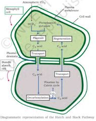
A) Part-I: Reactions in Granal chloroplast of Mesophyll cells:-

a) Carboxylation (Primary CO2 fixation):-
Atmospheric CO2 is accepted by phospho-enol pyruvic acid (PEPA) and get converted in oxalo acetic acid (OAA), a four-carbon compound in the presence of enzyme PEP carboxylase.
b) Reduction:- Oxalo-acetic acid is reduced to malic acid in the presence of NADPH2 and an enzyme malate dehydrogenase (or it get converted into aspartic acid by amination in the presence of NADPH2 and an enzyme transaminase).
Malic acid (or aspartic acid) is then transported in to chloroplasts of bundle sheath cells.
Part-Il: Reactions in Agranal chloroplast of Bundle sheath cells:-
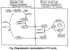
a) Decarboxylation:-
In these chloroplasts, malic acid undergoes oxidative decarboxylation in the presence of NADP to form pyruvic acid and CO2 is released. (If aspartic acid is formed, it undergoes deamination to form pyruvic acid).
The CO2 released is accepted by a second CO2 acceptor, i.e., RUBP.
Later these CO2 are fixed by C-3 pathway (Calvin cycle).
The 3 carbon molecule, i.e., Pyruvic acid is transported back to mesophyll cells. It is then phosphorylated by ATP to form PEPA. Initial acceptor is thus regenerated to continue the pathway.
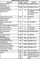
Difference between C3 and C4 plant.
Crassulacean Acid Metabolism:
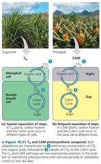
1) In certain plants, (members of family Crassulaceae) like succulent xerophytes, stomata remain closed during day (scotoactive stomata) to check the loss of water due to transpiration.
2) In these plants, gaseous exchange takes place during night time when stomata open.
3) During night time CO2 is available for fixation but light is unavailable. During daytime light energy is available (ATP and NADPH2 become available) but CO2 is not available due to complete closure of stomata.
4) Therefore, to overcome these difficulties these plants evolved alternate CO2 fixationpathway.
5) Mechanism of glucose synthesis in these plants is called Crassulacean acid metabolism.
6) Such plants are called as CAM plants. Kranz anatomy is absent in CAM plants.

In these plants, CO2 enters during night, when stomata are open.
It is taken up by PEPA (primary acceptor of CO2 ) and Oxaloacetic acid is formed in the presence of enzyme PEPcarboxylase.
Source of PEPA in CAM plants is starch.
) Oxaloacetic acid then gets reduced to malic acid in the presence of enzyme, malatede hydrogenase.
) Malate gets accumulated during night. This accumulation of malic acid is called as acidification. It takes place during night time.
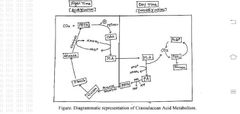
During day, when stomata are closed, malate undergoes oxidative decarboxylation gradually and gets converted into pyruvate. This oxidative decarboxylation of malic acid is called as deacidification.
) CO2 enters in Calvin cycle and sugar is produced.
) Pyruvate also gets converted into starch.
) During night time, again PEPA is regenerated from starch.
) Acid concentration increases during night and decreases during day.
) This diurnal (day and night) fluctuation in acid concentration is characteristic feature of CAM plants.
Opuntia, Euphorbia, 🌵 Cactus, Pineapple, Bryophyllum
Factors affecting photosynthesis:
Photosynthesis is under the influence of several factors, both (plant) internal andexternal (environmental).
The plant factors include the number, size, age and orientation of leaves, mesophyll cells and chloroplasts, internal CO2 concentration and the amount of chlorophyll. The plant or internal factors are dependent on the genetic predisposition and the growth of the plant.
) The external factors would include the availability of sunlight, temperature, CO2 concentration and water.
Blackman’s (1905) Law of Limiting Factors
When several factors affect any [bio] chemical process, Blackman’s (1905) Law of Limiting Factors comes into effect.
This states the following:
If a chemical process is affected by more than one factor, then its rate will be determined by the factor which is nearest to its minimal value: it is the factor which directly affects the process if its quantity is changed.
) For example, despite the presence of a green leaf and optimal light and CO2 conditions, the plant may not photosynthesize if the temperature is very low. This leaf, if given the optimal temperature, will start photosynthesizing.
) As a plant photosynthesizes, all these factors will simultaneously affect its rate. Hence, though several factors interact and simultaneously affect photosynthesis or CO2 fixation, usually one factor is the major cause or is the one that limits the rate. Hence, at any point the rate will be determined by the factor available at sub-optimal levels.
Effect of Light on photosynthesis:
light quality
light intensity
light duration
Light quality:
Light between 400 – 700 nm wavelength constitute the photosynthetically active radiation (PAR). Maximum photosynthesis takes place in red and blue light of visible spectrum and minimum photosynthesis takes place in green light
Light intensity: There is a linear relationship between incident light and CO2 fixation rates at low light intensities. At higher light intensities, gradually the rate does not show further increase as other factors become limiting (Figure -------). 4) What is interesting to note is that light saturation occurs at 10 per cent of the full sunlight. Hence, except for plants in shade or in dense forests, light is rarely a limiting factor in nature. 5) Increase incident light beyond a point causes the breakdown (photo-oxidation or solarization) of chlorophyll and a decrease in photosynthesis. 6) C4 plants have high light saturation point than C3 plants.
Duration of light never affects rate of photosynthesis (if quality/ wavelength and intensity of light are constant).
) Although light is rarely act as a limiting factor in photosynthesis but under following circumstances light acts as a limiting factor:
Plants in shade
In dense forest
Cloudy sky
In deep sea plants
Light compensation point:
At specific light intensity
There is no net atmospheric gaseous exchange.
Rate of photosynthesis is equal to the rate of respiration .
There is no net carbon assimilation or carbon fixation.
There is no net glucose synthesis.
There is no net productivity.
Effect of Carbon dioxide concentration on photosynthesis
Carbon dioxide is the major limiting factor for photosynthesis. 2) The concentration of CO2 is very low in the atmosphere (between 0.03 and 0.04 per cent or 300 to 400 ppm). 3) Increase in concentration up to 0.05 per cent can cause an increase in CO2 fixation rates; beyond this the levels can become damaging over longer periods. 4) The C3 and C4 plants respond differently to CO2 concentrations. 5) At low light conditions neither group responds to high CO2 conditions. At high light intensities, both C3 and C4 plants show increase in the rates of photosynthesis.
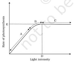
Based on the graph, answer the following questions. (a) At which point/s (A, B or C) in the curve is light a limiting factor?
(b) What could be the limiting factor/s in region A?
(c) What do C and D represent on the curve?
(a)
Ans. At regions A and B light is the limiting factor.
(b)Ans. In the region ’A’, light can be a limiting factor.
(c) Ans. ‘C’ is the region where the rate of the photosynthesis is not increased when light intensity is increased. D is the point where some other factors become limiting.
What is important to note is that the C4 plants show saturation at about 360 µlL-1 (360 ppm or 0.036 per cent) while C3 responds to increased CO2 concentration and saturation is seen only beyond 450 µlL-1 (450 ppm or 0.045 per cent).
Thus, current availability of CO2 levels is limiting to the C3 plants. 8) The fact that C3 plants respond to higher CO2 concentration by showing increased rates of photosynthesis leading to higher productivity (CO2 fertilization effect) has been used for some greenhouse crops such as tomatoes and bell pepper. They are allowed to grow in carbon dioxide enriched atmosphere that leads to higher yields.
At specific CO2 concentration There is no net atmospheric gaseous exchange. Rate of photosynthesis is equal to the rate of respiration. There is no net carbon assimilation or carbon fixation. There is no net glucose synthesis. There is no net productivity. CO2 compensation point for C4 plants is 0 to 10 ppm. CO2 compensation point for C3 plants is 25 to 100 ppm
Effect of Temperature on photosynthesis:
The dark reactions being enzymatic are temperature controlled. 2) Though the light reactions are also temperature sensitive they are affected to a much lesser extent. 3) The C4 plants respond to higher temperatures (30 to 400C) and show higher rate of photosynthesis while C3 plants have a much lower temperature optimum (20 to 250C)
The temperature optimum for photosynthesis of different plants also depends on the habitat that they are adapted to. Tropical plants have a higher temperature optimum than the plants adapted to temperate climates.
Effect of Water on photosynthesis:
1) Even though water is one of the reactants in the light reaction, the effect of water as a factor is more through its effect on the plant, rather than directly on photosynthesis. 2) Water stress causes the stomata to close hence reducing the CO2 availability. Besides, water stress also makes leaves wilt, thus, reducing the surface area of the leaves and their metabolic activity as well.
Inhibitors of Photosynthesis:
Inhibitors are used as weedicides or herbicides. Inhibitors that block PS II and stop non-cyclic photophosphorylation. [1] DCMU (Dichlorophenyl Dimethyl urea)/ Diuron. [2] CMU (Chlorophenyl Dimethyl Urea)/ Monouron. [3] PAN (Peroxy Acetyl Nitrate). Inhibitors that block PS I and stop cyclic photophosphorylation. [1] Diquat [2] Paraquat


Comments
Post a Comment